Monte Carlo (MC) Studies of Radiation Transport
Development of Monte Carlo (MC) Dose Engine
Big Data and Machine Learning Techniques in Radiation Oncology
Cancer Prediction and Risk Estimation Through Artificial Intelligence
Digital Health
Collaborators
Monte Carlo (MC) Studies of Radiation Transport |
Go to the Top |
MC simulations are very helpful in studying radiation transport in human body. Through this method, one can predict many quantities of interest that are difficult to measure physically. For example, to study radiation dosimetry, in-vivo dosimetry, planning radiotherapy in patients, estimation of radiation doses in sensitive organs during radiotherapy, radiation protection in radionuclide therapy etc. These are also helpful in nuclear medicine and radiology for estimating absorbed doses during different imaging studies. Several general-purpose MC based computer codes are currently available for the radiation transport at our lab (i.e., EGSnrc, GEANT4, TOPAS). These codes are used for different studies related to photon therapy and particle therapy (see figures). The simulations studies normally require validation with experimental data. For experimental part of the research, our lab has active collaborations with various cancer clinics nearby such as South Florida Proton Institute (SFPTI), Lynn Cancer Center at Boca Rational Hospital and GenesisCare, Florida.
|
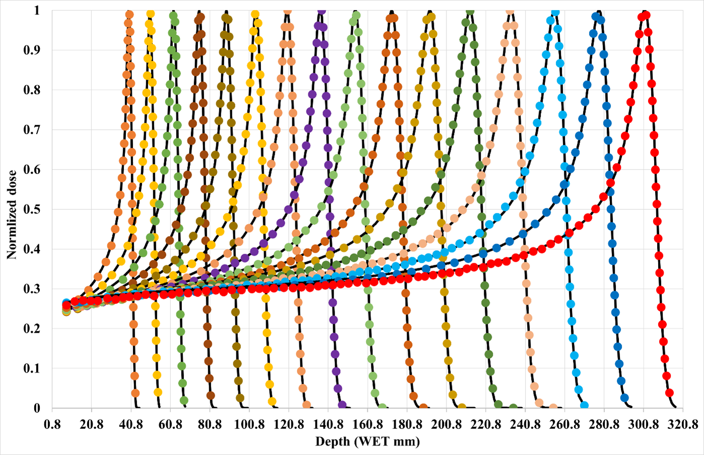
| Figure. Experimental (black lined) and simulation (dashed colorful lines) IDDs for 70-220 MeV proton energies from left to right with 10 MeV energy step. |
| Figure: A three-dimensional view of a treatment plan to treat a very small tumor in left trigeminal nerve using a fixed virtual cone technique. The fixed virtual cone is characterized by 0.5 cm x 0.5 cm high-definition (HD) MLC field, defined by two pairs of central leaves of the HDMLC. |
 MLC field, defined by two pairs of central leaves of the HDMLC.")
|

|
| Figure: Comparison of two-dimensional dose profiles obtained from (a) MC simulation and (b) TPS using the MapCHECK QA set up (SSD = 730 mm, z = 70 mm) for 15.4 mm x 15.4 MLC field with 1 mm voxel size in water phantom. |
Development of Monte Carlo (MC) Dose Engine |
Go to the Top |
The Monte Carlo (MC) method is widely used to solve various problems in radiation and medical physics. However, computational efficiency has been a main concern and therefore, limiting its applications in the clinical environment. Recently, graphics processing unit (GPU) drew the attention of relevant computational scientists working in the field of medical physics due to its effective scientific computing capability (i.e., rapid parallel processing capability). Its capability is really tested with graphic cards (e.g., NVIDIA’s GeForce and Tesla series) in the field of medical physics and medical image processing. Our aim is to develop a fast GPU based MC dose engine (see, figures). The aim is to improve the computational efficiency as compared with conventional CPU-based calculations. The dose engine will be used to estimate the doses received by the patients during different diagnostic and radiotherapy procedures. The accuracy and efficiency of the developed GPU-based MC code will be tested with different clinical cases (e.g., a 3D conformal radiotherapy of breast cancer, image-guided IMRT treatments of brain, head and neck, prostate and spine lesions, and an image-guided VMAT treatment of lung cancer etc.).
|
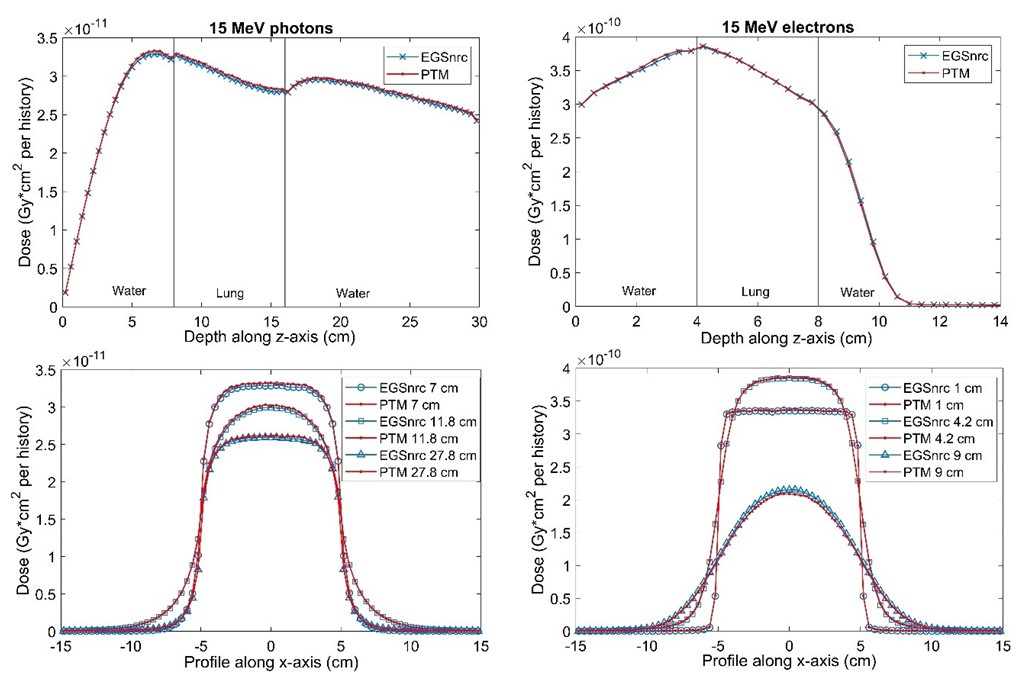
| PDD & lateral profiles at different depths (bottom row) in water-lung-water multilayered phantom |
|

|
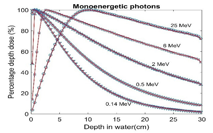
| PDD in a water phantom. Our MC code results are shown in red lines with dots while EGSnrc results are shown in blue lines with open symbols |
Big Data and Machine Learning Techniques in Radiation Oncology |
Go to the Top |
Artificial intelligence (AI) and machine learning (ML) approaches have caught the attention of many in health care scientist. Current literature suggests there are many potential benefits that could transform future clinical workflows and decision making. Embedding AI and ML concepts in radiation oncology could be a fundamental step in equipping radiation professional to engage in competent and safe practice as they utilize clinical technologies. Therefore, the goal is to develop a functional, dynamic, deep reinforcement learning (DRL) model to determine an optimal treatment strategy for each cancer patient. It will consider relevant parameters for a patient's health, clinical conditions (i.e., patient-specific data), and individual patient’s tumor characteristics and dynamics as well as prior oncological treatment outcomes for similar patients. Our main targets are:


Cancer Prediction and Risk Estimation Through Artificial Intelligence |
Go to the Top |
Early detection of a cancer is a challenging task because of either advanced stage occurrence of cancer-specific symptoms and/or lack of a reliable screening tool to identify high-risk patients. If the screening tools are available, the challenge is screening a large population. Although, a lot of health data is generated daily through electronic medical records from primary care centers in US however, there is no tool available to standardize and analyze this data collectively. If this data is properly managed and analyze, the increase in the cancer specific risk factors for different kind of cancers is expected. Our aim is to develop a multi-parameterized artificial neural network based on easily available personal health data. The network will be used predict cancer with high sensitivity prior to symptom onset and stratify the large population in term of cancer risk.

Digital Health |
Go to the Top |
With the increased digitization in healthcare, immense amount of data is generated from different segments of healthcare industry other than hospitals and healthcare providers, for example, social media platforms, medical insurance, medical equipment, life sciences and medical research. There is little academic knowledge regarding how information technology (IT) can be applied to the data to transform conventional healthcare into digital health through new era of AI and Big Data analytics. By filling this research gap, our aim is to successfully apply Machine intelligence algorithms to medical data in order to revolutionize several aspects of medical practice by enhancing diagnosis accuracy, diagnosing the source and progression of disease, as well as creating viable treatments for cancer diseases.

Collaborators |
Go to the Top |
